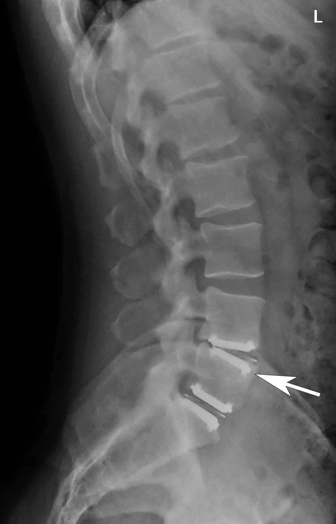

#Herniated disk xray images movie
The dye highlights the tissues to be evaluated and shows a continuous x-ray movie on a computer screen.

This process is called fluoroscopy and includes the injection of a contrast agent or dye into the target tissue. Physicians may use x-rays for live guidance while evaluating internal spinal structures, such as the spinal joints or nerves. Computerized Tomography (CT) Scan with Myelogram.Introduction to Diagnostic Studies for Back and Neck Pain.A tumor, which is also a mass of tissue, can be visualized on an x-ray due to its bulk and position. Similarly, neural tissues, like the spinal cord and spinal nerves, that may cause back or neck pain are usually not well defined in conventional x-rays. Muscles, ligaments, and tendons, which are often a source of back and neck pain, are not very well-visualized on a conventional x-ray. Soft tissues allow more x-ray to pass through them, creating a black impression on the photographic plate. The structure, shape, and alignment of the spinal vertebrae and facet joints can be well-documented in spinal x-rays.

Bones are the best and most clearly visualized structures on an x-ray. X-ray of spinal bonesĭense tissues with high calcium content, such as bone, absorb less x-ray and create a white-colored image on the photographic film. Spinal x-rays can be captured to specifically focus on the 3 main parts of the spine– cervical spine (neck), thoracic spine (mid-back), and lumbar spine (lower back) as well as the pelvis, which includes the sacroiliac joint. 3 When they pass through the tissues, x-rays have the ability to create two-dimensional black-and-white images of the structure(s) that they’ve traversed on a photographic film placed on the opposite side of the body. X-rays are high-frequency energy waves that can penetrate through the body and are either absorbed, reflected, or traversed through the target tissue(s).


 0 kommentar(er)
